Steinberg Lab Research: Stem Cells
STEM CELL THERAPY offers enormous promise for the majority of the 795,000 Americans yearly who suffer a stroke yet currently have no pharmacological therapy to promote recovery. Preclinical data from our lab and others have demonstrated that stem cell transplantation can enhance stroke recovery. This has led to major efforts to advance stem cell therapy for stroke to the clinic, including our human neural stem cell (hNSC) product, NR1 cells, which is transitioning to the clinic via a California Institute for Regenerative Medicine (CIRM)-funded Disease Team program.
Understanding how hNSCs interact with the stroke-injured brain to induce recovery is crucial to maximizing their effectiveness. Once we understand the mechanisms by which transplanted hNSCs exert therapeutic effects, we can exploit their full clinical potential as well as predict and prevent potential side effects.
Consequently, we are studying optimal parameters for successful transplantation strategies in rodent models of stroke; these parameters will depend on the cross talk between the graft and the host and will facilitate understanding of the mechanisms underlying cell-enhanced functional recovery.
Stroke damage encompasses a wide range of pathologies. The initial vascular damage of stroke triggers a cascade of damaging events that occur days after the initial infarct. The unique advantage of using stem cells for post-stroke recovery lies in the multiple modalities through which they enhance recovery.
Our current paradigm is that hNSCs transplanted into the host stroke brain, rather than exerting their effects directly by replacing damaged tissue, secrete factors and interact with the stroke milieu in a manner that stimulates endogenous repair mechanisms that are activated by stroke.
We have found (eg., Horie et al Stem Cells 2011) that transplanted hNSCs can attenuate some of the direct effects of stroke damage, such as inflammation and vascular leakage1. These effects are mediated in part by vascular endothelial growth factor (VEGF). Strikingly, VEGF produced by transplanted hNSCs (human central nervous system stem cells grown as neurospheres or hCNS-SCns) also enhanced the endogenous repair mechanism of vascular regeneration (Figure 1), which supports our idea that transplanted stem cells enhance host repair mechanisms to promote functional recovery.

Figure 1 (A) hCNS-SCns (red: human cytoplasmic marker SC121) survive and migrate over time (green: lectin-positive blood vessels; blue: DAPI). * indicates lesion. Inset shows higher magnification of hCNS-SCns. Scale bar = 100 μm (50 μm in inset, except 3 week inset: 25 μm). (B): hCNS-SCns-treated animals compared with buffer-treated animals show significantly improved behavioral recovery after stroke 1.
Neuroplasticity is another endogenous repair mechanism that occurs during functional recovery. Using axonal and dendritic tracing methods we found that hNSCs enhance neuronal structural plasticity by increasing dendritic branching and axonal transport. In vitro, functional assays identified several hNSC-secreted factors that can increase neurite sprouting (eg., Steinberg et al Brain 2011).
Building on this observation that transplanted hNSCs can induce structural plasticity in the post-stroke brain we are now focusing on changes that can be induced at the synaptic level. Our approach integrates the advanced proteomic imaging technique, array tomography, to accurately identify and count excitatory and inhibitory synapses (Figures 2 & 3), and electrophysiology techniques to measure functional changes at the synaptic and circuit level.
Inflammation plays a pivotal role in the extent of brain damage and recovery after a stroke. We showed at a rudimentary level that hNSCs reduce the number of microglia/macrophage present in the brain after stroke (1). We are now interested in how the hNSCs alter inflammation, what sub-populations of immune cells are affected, and how this relates to hNSC-induced recovery after stroke.
References
- Horie N, Niizuma K, Pereira MP, Sun GH, Keren-Gill H, Encarnacion A, Shamloo M, Hamilton SA, Jiang K, Huhn S, Palmer T, Bliss TM, Steinberg GK: Transplanted stem cell-secreted VEGF effects post-stroke recovery, inflammation, and vascular repair. Stem Cells 29(2):274-285, 2011
- Andres RH, Horie N, Slikker W, Keren-Gill H, Zhan K, Sun GH, Sheikh LA, McMillan EL, Schaar BT, Svendsen CN, Bliss TM, Steinberg GK: Human neural stem cells enhance structural plasticity and axonal transport in the ischemic brain. Brain 134(Pt 6): 1777–1789, 2011.
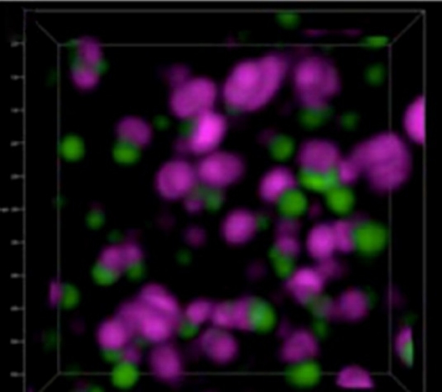
Figure 2. Pre- and post-synaptic marker colocalization as seen with array tomography, an imaging method co-invented by Stephen Smith, PhD and Kristina Micheva, PhD at Stanford. This 3-D reconstruction of a small volume (6 x 6 x 2 µm) shows the pre-synaptic marker synapsin (magenta) and the post-synaptic marker of glutamatergic synapses PSD95 (green). Individual synapses are well resolved. With permission from Dr. Stephen Smith.
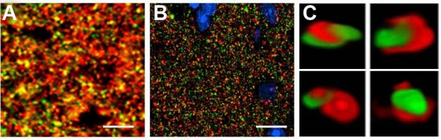
Figure 3. Differences in synaptic resolution by (A) confocal microscopy, and (B, C) array tomography. (C) 3D reconstruction of colocalized puncta. Red: pre-synaptic marker synapsin; Green: post-synaptic marker PSD95. Scale bar = 10 µm. With permission from Dr. Stephen Smith.
References
- Horie N, Niizuma K, Pereira MP, Sun GH, Keren-Gill H, Encarnacion A, Shamloo M, Hamilton SA, Jiang K, Huhn S, Palmer T, Bliss TM, Steinberg GK: Transplanted stem cell-secreted VEGF effects post-stroke recovery, inflammation, and vascular repair. Stem Cells 29(2):274-285, 2011
- Andres RH, Horie N, Slikker W, Keren-Gill H, Zhan K, Sun GH, Sheikh LA, McMillan EL, Schaar BT, Svendsen CN, Bliss TM, Steinberg GK: Human neural stem cells enhance structural plasticity and axonal transport in the ischemic brain. Brain 134(Pt 6): 1777–1789, 2011.
Banner photos Left: Transplanted stem cells (green) migrating towards blood vessels in stroke-damaged brain. Right: Human neural progenitor cells (red) are found in close proximity to blood vessels (green).